What is diabetic retinopathy?
Diabetic retinopathy is a most common diabetic eye disease caused by changes in the blood vessels of the retina which is the light-sensitive tissue at the back of the eye. The abnormal new blood vessels grow on the surface of the retina while the blood vessels of people with diabetic retinopathy may leak blood and grow fragile.
Diabetic retinopathy is a leading cause of low vision and blindness in many countries.
Two types of diabetic retinopathy
1. Background or nonproliferative diabetic retinopathy (NPDR)
It has mild, moderate and severe stages which can cause changes in the eye, including:
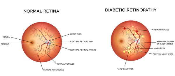
- Microaneurysms
- Small bulges in blood vessels of the retina that often leak fluid.
- Retinal hemorrhages
- Tiny spots of blood that leak into the retina.
- Hard exudates
- Deposits of cholesterol or other fats from the blood that have leaked into the retina.
- Macular edema
- Swelling or thickening of the macula caused by fluid leaking from the retina's blood vessels. The macula doesn't function properly when it is swollen. Macular edema is the most common cause of vision loss in diabetes.
- Macular ischemia
- Small blood vessels (capillaries) close. Your vision blurs because the macula no longer receives enough blood to work properly.
2. Proliferative diabetic retinopathy (PDR)
PDR may cause more severe vision loss than NPDR because it can affect both central and peripheral vision. PDR affects vision in the following ways:
- Vitreous hemorrhage
- Delicate new blood vessels bleed into the vitreous — the gel in the center of the eye — preventing light rays from reaching the retina. If the vitreous hemorrhage is small, you may see a few new, dark floaters. A very large hemorrhage might block out all vision, allowing you to perceive only light and dark. Vitreous hemorrhage alone does not cause permanent vision loss. When the blood clears, your vision may return to its former level unless the macula has been damaged.
- Traction retinal detachment
- Scar tissue from neovascularization shrinks, causing the retina to wrinkle and pull from its normal position. Macular wrinkling can distort your vision. More severe vision loss can occur if the macula or large areas of the retina are detached.
- Neovascular glaucoma
- If a number of retinal vessels are closed, neovascularization can occur in the iris (the colored part of the eye). In this condition, the new blood vessels may block the normal flow of fluid out of the eye. Pressure builds up in the eye, a particularly severe condition that causes damage to the optic nerve.
Symptoms
- Blurry vision
- Rings around lights
- Dark spots or flashing floaters
- Sudden loss of vision
- Leaking fluid
Often there are no symptoms in the early stages. You'd better contact your ophthalmologist for a complete exam if you are experience one or more of above symptoms.
Risk Factors
- High blood pressure
- Two type of diabetes
- Pregnant woman with diabetes
- Sleep apnea
- Elevated blood cholesterol levels
Prevent or treatment of diabetic retinopathy
Regular eye exam, strict control blood sugar and pressure levels are greatly helpful to reduce or prevent vision loss from diabetic retinopathy. Unless you have macular edema, the treatment is not necessary. And until now, there is no treatment to cure diabetic retinopathy. Contact your eye care professional for specific suggestions if you have vision loss.